Home
/ Micrograph Of Animal Cell With Labels - Quotes About Animal Cell 22 Quotes - You should concentrate on the similarities in form that permit identification of the.
Micrograph Of Animal Cell With Labels - Quotes About Animal Cell 22 Quotes - You should concentrate on the similarities in form that permit identification of the.
Micrograph Of Animal Cell With Labels - Quotes About Animal Cell 22 Quotes - You should concentrate on the similarities in form that permit identification of the.. The animal cell is more fluid or elastic or malleable in structure; (2) they are spherical or irregular membrane bound vesicles filled with digestive enzymes. Make your work easier by using a label. Cell is a tiny structure and functional unit of a living organism containing various parts known as organelles. Animal cells need a small drop of iodine or methylene blue to be seen under the microscope, with a coverslip placed on top.
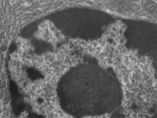
Transmission electron micrograph (tem) of lysosomes. Cytoplasm, plasma membrane, rough endoplasmic reticulum, golgi apparatus, lysosome, vacuole, starch granules, cell wall. The figure below is a fine structure of a generalized animal cell. Make your work easier by using a label. Drag the given words to the correct blanks to complete the labeling!
Unique Characteristics Of Eukaryotic Cells Microbiology from s3-us-west-2.amazonaws.com It's the cell's brain, employing chromosomes to instruct other parts of the cell. See the answer see the answer see the answer done loading. Cytoplasm complete the following chart: (3) d.) give one structural adaptation. Draw and label the detail of these cells as. Animal cells are packed with amazingly specialized structures. This is the electron micrograph of lysosome, and is characterized by following features. See how a generalized structure of an animal cell and plant cell look with labeled diagrams.
Drag the given words to the correct blanks to complete the labeling!
This is the electron micrograph of lysosome, and is characterized by following features. Electron micrograph of plant cell labeled anatomy and physiology of animals animal cell electron microscope wide collections of all kinds of labels pictures online. The electron micrograph of lysosomes: Nucleus plant cell microscope labeled. Cytoplasm, plasma membrane, rough endoplasmic reticulum, golgi apparatus, lysosome, vacuole, starch granules, cell wall. The cell is the basic unit of life. Cell wall, nucleus and chloroplasts can be seen with a compound light microscope under a total magnification of 400 x. For many years until the electron microscope was invented this was. Control center of the cell. This organelle is like the post office of the cell. (place an x in the appropriate box) Onion cell drawing (high power) 2. Link to a random quiz page.
Control center of the cell. Draw and label the detail of these cells as. See how a generalized structure of an animal cell and plant cell look with labeled diagrams. Horoplast, nucleus, nucleolus, chromatin, nuclear membrune, the micrograph above represents a lymphocyte. Mitochondria is a plural word.
Animal Plant Cells Cie A Level Biology Revision Notes from cdn.savemyexams.co.uk (place an x in the appropriate box) Nucleus plant cell microscope labeled. One vital part of an animal cell is the nucleus. You should concentrate on the similarities in form that permit identification of the. Animal cell diagram under microscope. Transmission electron micrograph (tem) of lysosomes. Cytoplasm complete the following chart: Here is an electron micrograph of an animal cell with the labels superimposed:
Bookfanatic89 diagram of plant cell under electron microscope.
It's the cell's brain, employing chromosomes to instruct other parts of the cell. Human (animal) cell under microscope (3d illustration) internal structure of an animal cell. Include plasma membrane, cytoplasm, nuclear envelope, nuclear pore, chromatin, rough er, smooth er, golgi body and mitochondrian. Below is a collection of electron micrographs with labelled subcellular structures that you should be able to identify. Cell is a tiny structure and functional unit of a living organism containing various parts known as organelles. Learn vocabulary, terms, and more with flashcards, games, and other study tools. (2) they are spherical or irregular membrane bound vesicles filled with digestive enzymes. Bookfanatic89 diagram of plant cell under electron microscope. Animal cells introduction background information: You see that many features are in common. Animal cells 13 february 2013 lesson description in this lesson we will discuss the following: Animal cell diagram under microscope. Plexus probio5 label march 26th 2018 | free labels wide collections of all kinds of labels pictures online.
As observed in the labeled animal cell diagram, the cell membrane forms the confining factor of the cell, that is it envelopes the cell constituents together and gives the cell its shape, form, and existence. Provide labels for parts a, b and c. See how a generalized structure of an animal cell and plant cell look with labeled diagrams. (place an x in the appropriate box) (2) they are spherical or irregular membrane bound vesicles filled with digestive enzymes.
Draw And Label Generalized Animal Cell Shefalitayal from o.quizlet.com A thin layer of elodea, an aquatic plant, works well for an example of a. The cell theory terminology parts of a microscope the definition of a cell examples of cells type of cells plant and animal cells parts of animal cells: Animal cells introduction background information: However, the cell membrane in plant cells is quite rigid, while, the cell membrane in animal cells is quite flexible. Cell wall, nucleus and chloroplasts can be seen with a compound light microscope under a total magnification of 400 x. Control center of the cell. All organisms are made up of cells (or in some cases, a single cell). Animal cells need a small drop of iodine or methylene blue to be seen under the microscope, with a coverslip placed on top.
See the answer see the answer see the answer done loading.
As observed in the labeled animal cell diagram, the cell membrane forms the confining factor of the cell, that is it envelopes the cell constituents together and gives the cell its shape, form, and existence. Maybe you would like to learn more about one of these? Cell is a tiny structure and functional unit of a living organism containing various parts known as organelles. We did not find results for: The animal cell is more fluid or elastic or malleable in structure; Cytoplasm complete the following chart: Animal cells 13 february 2013 lesson description in this lesson we will discuss the following: Horoplast, nucleus, nucleolus, chromatin, nuclear membrune, the micrograph above represents a lymphocyte. Start studying aice biology chapter 1: After hundreds of years of observations by many biologists, the cell theory was developed. Comparative illustration of plant and animal cell anatomy (with labels). Human (animal) cell under microscope (3d illustration) internal structure of an animal cell. However, the cell membrane in plant cells is quite rigid, while, the cell membrane in animal cells is quite flexible.
Share :
Post a Comment
for "Micrograph Of Animal Cell With Labels - Quotes About Animal Cell 22 Quotes - You should concentrate on the similarities in form that permit identification of the."


Post a Comment for "Micrograph Of Animal Cell With Labels - Quotes About Animal Cell 22 Quotes - You should concentrate on the similarities in form that permit identification of the."